
Overview of surgery
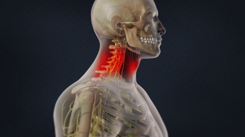
Posterior cervical spine surgery is performed from the back of the neck to relieve compression on the nerves and the spinal cord and to treat painful arthritis. This procedure is considered when the nerves and spinal cord are being compressed from the back or may occasionally be considered in combination with an anterior (front) surgery.
Steps of the surgery
- The surgery is performed under general anaesthesia, with the patient lying on the tummy (face down)
- The spine is exposed through an incision on the skin at the back of the neck. The spine is exposed by moving the muscles aside. The involved bones and ligaments are removed using special instruments and the pressure on the nerve roots and spinal cord is relieved
- If a fusion is planned, titanium screws will be inserted into the bones of the spine and connected with rods and supplemented with bone graft
- The incision (cut) is closed with dissolvable sutures. A drain tube will remove the blood that collects at the surgical site. A urinary tube may be used to drain the bladder.
Success rates and the outcomes:
This surgery is associated with a high success rate (>90%) in terms of relief of the arm pain. If muscle weakness and numbness was present prior to surgery, it usually improves over a period of time. If myelopathy (spinal cord dysfunction) was present before surgery, the extent of recovery is less predictable. However, surgical decompression will usually stop the deterioration and provides the most favourable opportunity for recovery. If the surgery has been performed for neck pain, relief of symptoms usually occurs over weeks to months.
Your pathway to recovery:
- Following surgery, the patient will be moved to the recovery room where a neck collar is provided to ensure comfort and to prevent excessive neck movements. Adequate pain relief and intravenous fluids will be provided. When the patient is comfortable, he/she will be transferred to a high dependency ward for an overnight stay for more personalized attention
- Most patients will be encouraged to walk the day after surgery with the neck collar after removing the urinary tube. Normal diet or a soft diet is usually advised either in the evening after the surgery or the next morning
- The duration of stay in the hospital may extend from 2-4 days depending on comfort and ability to walk and the surgeon will decide on the date of discharge
Complications
Allergic reactions to medications and conditions like pneumonia, stroke or heart attack, though rare and not directly caused by the surgical treatment, may have serious consequences.
Surgical complications can include bleeding, infection, spinal fluid leak, injury to the veins and arteries near the spine, injury to the spinal cord and nerves and its surrounding protective layer (dura). Injury to the spinal cord or the nerves during surgery can result in varying degrees of muscular paralysis, alteration of normal sensations and loss of bowel and bladder control which may be permanent or may recover over a period of time. An injury to the covering layers of the nerves (dura) can result in a leak of spinal fluid and may rarely require a repeat surgery. The bone graft may not heal and fuse the spine (non-union) and may in some instances require further surgery.
A 1-5% incidence of post-operative wound infection is possible despite antibiotics given before and after surgery. Superficial infections can be treated with antibiotics, while deep infections may require a wound wash-out under anaesthesia. The presence of an infection in any other region (urinary bladder, chest and skin) immediately prior to surgery, increases the risk of post-operative infection, so inform your surgeon about this.